A bone fracture (sometimes abbreviated FRX or Fx, Fx, or #) is a medical condition in which there is a partial or complete break in the continuity of the bone. In more severe cases, the bone may be broken into several pieces. A bone fracture may be the result of high force impact or stress, or a minimal trauma injury as a result of certain medical conditions that weaken the bones, such as osteoporosis, osteopenia, bone cancer, or osteogenesis imperfecta, where the fracture is then properly termed a pathologic fracture.
Signs and symptoms
Although bone tissue itself contains no nociceptors, bone fracture is painful for several reasons:
- Breaking in the continuity of the periosteum, with or without similar discontinuity in endosteum, as both contain multiple pain receptors.
- Edema and hematoma of nearby soft tissues caused by ruptured bone marrow evokes pressure pain.
- Involuntary muscle spasms trying to hold bone fragments in place.
Damage to adjacent structures such as nerves, muscles or blood vessels, spinal cord, and nerve roots (for spine fractures), or cranial contents (for skull fractures) may cause other specific signs and symptoms.
Complications
Some fractures may lead to serious complications including a condition known as compartment syndrome. If not treated, eventually, compartment syndrome may require amputation of the affected limb. Other complications may include non-union, where the fractured bone fails to heal or mal-union, where the fractured bone heals in a deformed manner. One form of malunion is the malrotation of a bone, which is especially common after femoral and tibial fractures.
Complications of fractures may be classified into three broad groups, depending upon their time of occurrence. These are as follows –
- Immediate complications – occurs at the time of the fracture.
- Early complications – occurring in the initial few days after the fracture.
- Late complications – occurring a long time after the fracture.

An old fracture with nonunion of the fracture fragments
| Immediate complications | Early complications | Late complications |
|---|---|---|
Systemic
|
Systemic
|
Imperfect union of the fracture
|
Local
|
Local
|
Others
|
Pathophysiology
The natural process of healing a fracture starts when the injured bone and surrounding tissues bleed, forming a fracture hematoma. The blood coagulates to form a blood clot situated between the broken fragments. Within a few days, blood vessels grow into the jelly-like matrix of the blood clot. The new blood vessels bring phagocytes to the area, which gradually removes the non-viable material. The blood vessels also bring fibroblasts in the walls of the vessels and these multiply and produce collagen fibres. In this way, the blood clot is replaced by a matrix of collagen. Collagen's rubbery consistency allows bone fragments to move only a small amount unless severe or persistent force is applied.
At this stage, some of the fibroblasts begin to lay down bone matrix in the form of collagen monomers. These monomers spontaneously assemble to form the bone matrix, for which bone crystals (calcium hydroxyapatite) are deposited in amongst, in the form of insoluble crystals. This mineralization of the collagen matrix stiffens it and transforms it into bone. In fact, bone is a mineralized collagen matrix; if the mineral is dissolved out of bone, it becomes rubbery. Healing bone callus on average is sufficiently mineralized to show up on X-ray within 6 weeks in adults and less in children. This initial "woven" bone does not have the strong mechanical properties of mature bone. By a process of remodelling, the woven bone is replaced by mature "lamellar" bone. The whole process may take up to 18 months, but in adults, the strength of the healing bone is usually 80% of normal by 3 months after the injury.
Several factors may help or hinder the bone healing process. For example, tobacco smoking hinders the process of bone healing, and adequate nutrition (including calcium intake) will help the bone healing process. Weight-bearing stress on bone, after the bone has healed sufficiently to bear the weight, also builds bone strength.
Although there are theoretical concerns about NSAIDs slowing the rate of healing, there is not enough evidence to warrant withholding the use of this type analgesic in simple fractures.
Effects of smoking
Smokers generally have lower bone density than non-smokers, so they have a much higher risk of fractures. There is also evidence that smoking delays bone healing.
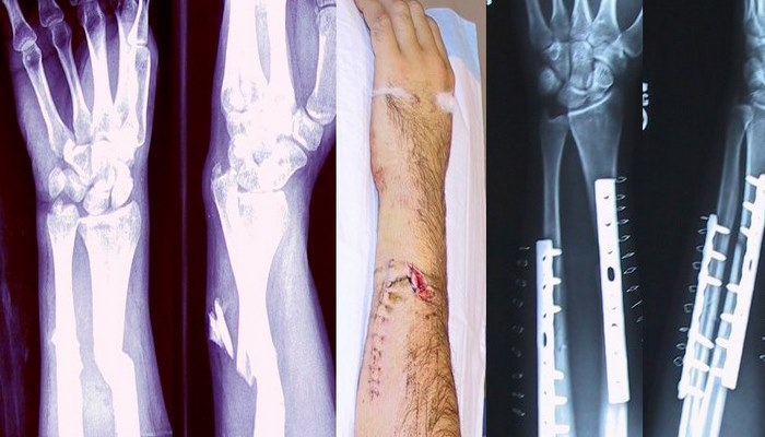
Diagnosis
A bone fracture may be diagnosed based on the history given and the physical examination performed. Radiographic imaging often is performed to confirm the diagnosis. Under certain circumstances, radiographic examination of the nearby joints is indicated in order to exclude dislocations and fracture-dislocations. In situations where projectional radiography alone is insufficient, Computed Tomography (CT) or Magnetic Resonance Imaging (MRI) may be indicated.